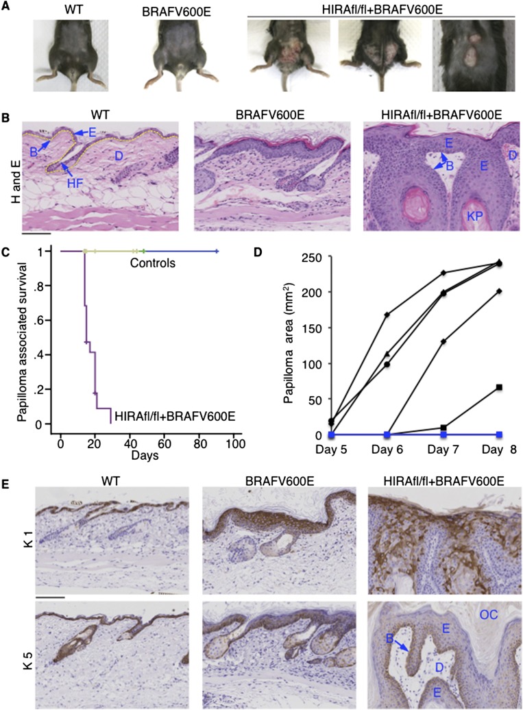
Figure 6.
HIRA suppresses oncogene-induced neoplasia. (A) Skin of representative wild-type (WT), AhCreER/LSL-BrafV600E, and AhCreER/LSL-BrafV600E/Hirafl.fl mice approximately 2 wk after tamoxifen treatment. (B) H+E-stained sections of skin from the indicated mice approximately 2 wk after tamoxifen treatment. (B) Basement membrane location (also indicated by dashed yellow line in wild type); (E) epidermis; (D) dermis; (HF) hair follicle; (KP) keratin pearl. (C) Papilloma-associated survival of control and AhCreER/LSL-BrafV600E/Hirafl.fl mice after tamoxifen treatment (N = 19; seven male, 12 female) for AhCreER/LSL-BrafV600E/Hirafl.fl mice. Controls include AhCreER/LSL-BrafV600E (N = 11; three male, eight female; light green), AhCreER/Hirafl.fl (N = 9; five male, four female; dark green), and AhCreER (N = 12; eight male, four female; blue). Mice culled of causes unrelated to papilloma formation were censored from analysis (indicated by “+” on the plot). See Supplemental Data Set 5 for all mouse outcomes. (D) Skin lesion area measured (length × width) at daily intervals after tamoxifen treatment. N = 5 for AhCreER/LSL-BrafV600E/Hirafl.fl (black); N = 2 for AhCreER/LSL-BrafV600E (blue), neither of which developed any skin lesion. In addition, AhCreER/LSL-BrafV600E (N = 2) and AhCreER/LSL-BrafV600E/Hirafl.fl (N = 6) were treated with corn oil and observed for 4 wk. None of the mice developed any lesions or tumors. (E) Skin from mice of the indicated genotypes ∼2 wk after tamoxifen treatment, stained with antibodies to keratin 1 and keratin 5. Histological features labeled as in B. (OC) Outer cornified layer. Bars, 100 μM.

