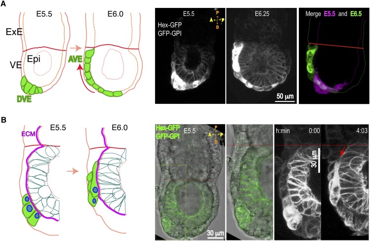
Figure 1.
AVE collective migration in the early mouse embryo. Transgenic embryos (E5.5–E6.5) expressing the indicated transgenes were imaged by DIC and confocal time-lapse microscopy. (A) Sagittal sections showing AVE (Hex-GFP) cells migrating over epiblast (GFP-GPI) cells. Schematic (left) and maximal projections (right) of four optical sections (6-μm thickness) taken from time-lapse snaphots (from Supplemental Movie S1). (Epi) Epiblast; (DVE) distal VE. A morphologically distinct, highly columnar subpopulation of VE cells expressing the Hex-GFP marker (green) and located at the distal end (DVE) move on the presumptive anterior side (red arrow), arrive at the embryonic/ExE border (red line), and become AVE. (B) Schematic (left) and optical projections (right) from time-lapse snapshots (10-μm thickness) of migrating Hex-GFP cells (green) moving on the ECM (magenta line). Long protrusions intercalate between front VE cells and the epiblast (red arrow).

