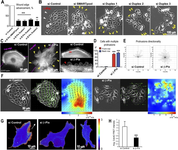
Figure 3.
β-Pix is required for collective epithelial migration. 16HBE epitheliocytes were used in an in vitro collective epithelial migration assay as cells in either a scratched monolayer or discrete islands. An siRNA screen of the 82 mammalian Rho family GEFs performed in the scratch assay identified β-Pix as a candidate regulator of collective migration. (A) siRNA-mediated depletion of β-Pix by an siRNA SMARTpool (mixture of four siRNAs) or the four individual duplexes inhibits wound edge advancement in a monolayer scratch assay (mean ± SEM; n = 3 independent experiments; unpaired Student’s t-test, [***] P < 0.001 for duplexes 1 and 2 and SMARTpool; [**] P = 0.01 for duplex 3). (B) Phase contrast images of 16HBE cells showing multiple protrusions at the free edges of cells all around epithelial islands (yellow arrows) in β-Pix-depleted cells. In control islands, protrusion distribution is polarized (white arrows) in the direction of migration. (C) Mosaic expression of EYFP-actin in front-row cells of a scratched monolayer reveals multiple protrusions at the free edge (pink dotted line) and at the posterior and lateral sides (yellow arrows) in β-Pix-depleted cells (live epifluorescence imaging). In control cells, a single lamellipodium can be seen at the free edge, with relatively quiescent rear edges. The prominent marginal bundle (MB) seen in control cells is disassembled in β-Pix-depleted cells (inset; red arrow). (D) Quantification showing that β-Pix depletion generates multiple protrusions in both front- and back-row cells (mean ± SEM; n = 3 independent experiments; unpaired Student’s t-test, [***] P < 0.001 for both front- and back-row cells). (E) Quantification of protrusion directionality shows a random distribution after β-Pix depletion (n = 60 control protrusions; n = 164 protrusions in β-Pix-depleted cells; three independent experiments). (F) Quantitative PIV analysis was used to analyze cell flow in migrating islands. A collectively migrating control island shows unidirectional vectors (middle image) and a gradient of velocities (heat map) (taken from Supplemental Movie S9). β-Pix-depleted islands lose both directionality of migration (middle image) and a collective gradient of velocities (heat map). (G) Maximum projections of confocal sections of migrating 16HBE cells transiently expressing a dual-chain Cdc42 biosensor. Fluorescent cells are surrounded by nonfluorescent cells. Active Cdc42 is located at the leading edge in a control cell, in contrast to reduced Cdc42 activity in β-Pix knockdown cells. The white arrow shows the direction of migration. (H) Quantification of Cdc42 FRET intensity in lamellipodial regions. Data are mean ± SD; 20 control cells and 25 siRNA duplex 1 β-Pix knockdown cells; unpaired Student’s t-test, (***) P < 0.0001.

