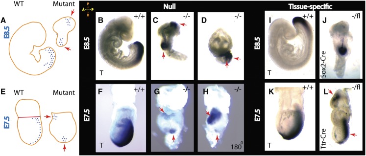
Figure 5.
Deletion of β-Pix results in anterior–posterior axis duplication. In situ hybridization analysis of Brachyury (T) expression is shown schematically at E8.5 (A) and E7.5 (E) for wild-type and β-Pix-null embryos. (B–D) Whole-mount in situ hybridization of wild-type littermate control embryos (β-Pix+/+) and β-Pix−/−-null (tm1b allele) embryos at E8.5. The primitive streak marker Brachyury (T) is seen on the posterior side of the wild-type embryo, while in β-Pix−/−-null embryos, Brachyury (T) is localized in two distinct sites (red arrows), thus duplicating the anterior–posterior axis. (F–H) At E7.5, the primitive streak marker in mutant embryos is located at two different sites (snapshots of the same embryo rotated 180°). (I–L) Tissue-specific knockouts reveal axis duplication after β-Pix knockout in VE cells (Ttr-Cre; β-Pix−/VE-deleted) (L) but not after β-Pix knockout in epiblast cells (Sox2-Cre; β-Pix−/epiblast-deleted) (J). At E6.5, a Brachyury (T) signal is not clearly established (see Supplemental Fig. S3E).

