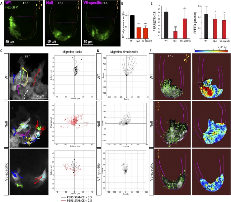
Figure 6.
Loss of β-Pix disrupts collective AVE migration. (A) An image from live confocal microscopy reveals AVE (Hex-GFP) cells located at the anterior side of control embryos at E6.5. AVE cells fail to migrate in β-Pix−/− (null) and β-Pix−/VE-deleted (VE-specific) mutant embryos and remain distal. (B) Quantification of AVE edge advancement toward the ExE border at E6.5 (mean ± SEM; n = 6 embryos; unpaired Student’s t-test, [***] P < 0.001 for both genotypes). (C) Cell tracking from time-lapse microscopy reveals disorganized migration of Hex-GFP cells in β-Pix−/− (null) and β-Pix−/VE-deleted (VE-specific) mutant embryos compared with wild type (from Supplemental Movie S10). (D) Quantification of migration directionality was performed in two independent experiments (n = 17 wild-type], 22 null, and 14 VE-specific cells per condition). (E) Quantification of AVE cell migration persistence (n = 20 cells in two null embryos; n = 17 cells in two wild-type embryos) and migration speeds (β-Pix−/−-null embryos: n = 20 cells, two embryos; β-Pix−/VE-deleted embryos: n = 15 cells, two embryos; wild-type embryos: n = 17 cells, two embryos). (F) PIV analysis of Hex-GFP migrating cells. Representative of three independent experiments. The left panels represent directionality of vectors, and the right panels show the spatial distribution of instantaneous velocities (heat map, meters per second). Pink lines outline the embryo and VE.

