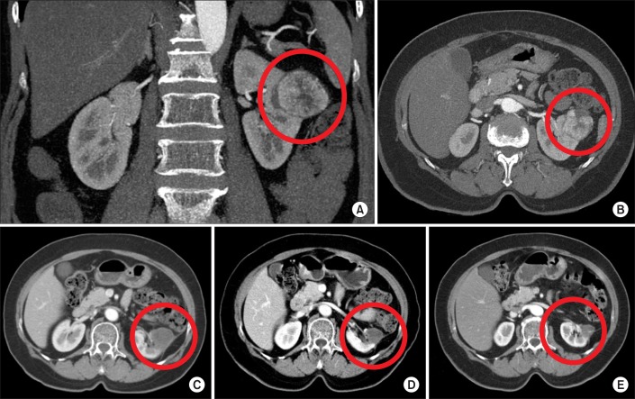
FIG. 2.
A 76-year-old female patient with a 3.9-cm left renal cell carcinoma (RCC) had a history of recent acute myocardial infarction, diabetes mellitus, hypertension, and high American Society of Anesthesiologists score. The figure shows the preoperative computed tomography (CT) scan (A and B) and the decreased size of the treated RCC in the left kidney without a definite viable portion at 3 months (C), 9 months (D), and 36 months (E) after left renal cryoablation by abdominal CT scan.

