
Fig. 2.
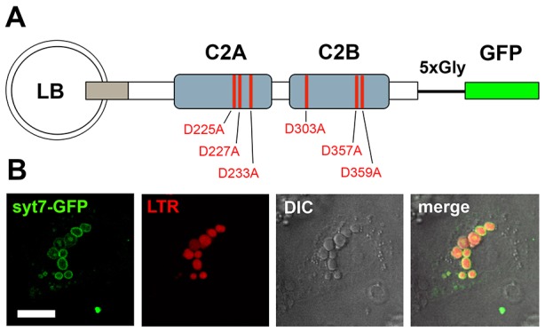
Syt7–GFP localises to the membrane of lamellar bodies. (A) Schematic representation of the Syt7–GFP constructs used in this study. Syt7 and GFP are separated by a short glycine linker. Red lines indicate amino acid positions in the Ca2+-binding sites in the two C2 domains (C2A and C2B) that were mutated to obtain Syt7 constructs with altered Ca2+-binding properties. (B) Syt7–EGFP (green) expressed for 24 h in ATII cells is primarily localised on the limiting membrane of lamellar bodies, as confirmed by co-staining of lamellar bodies with LTR (red). Scale bar: 5 µm.