
Fig. 3.
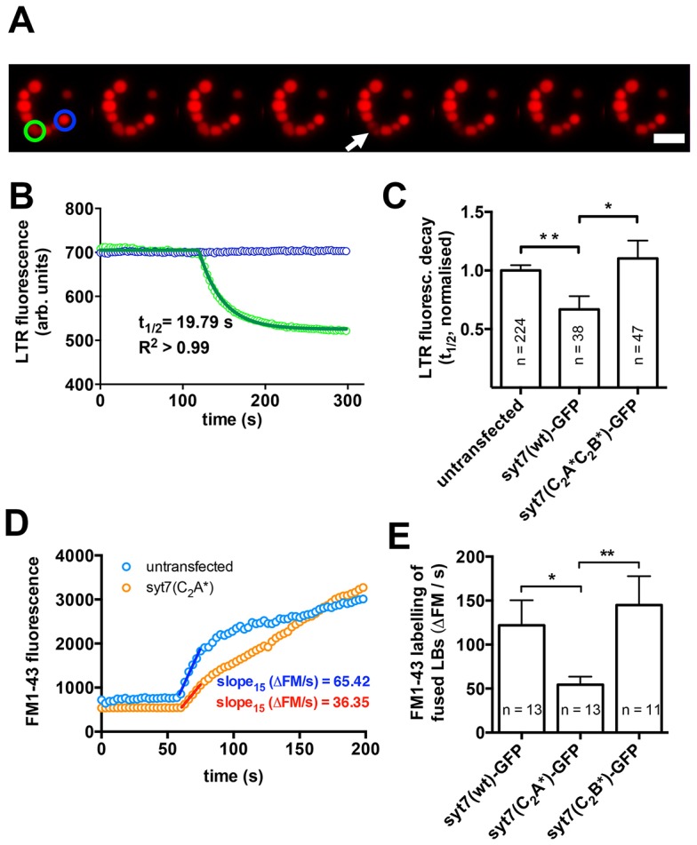
Syt7 facilitates fusion pore expansion following exocytic fusion of lamellar bodies with the plasma membrane. (A) Image sequence illustrating loss of LTR from an individual lamellar body following fusion with the plasma membrane and fusion pore opening (green circle). Note that the LTR fluorescence of a non-fusing vesicle (blue circle) does not change significantly. Scale bar: 5 µm. (B) Half-times (t1/2) of the fluorescence decay were analysed to compare diffusion of LTR across the fusion pore for various experimental conditions and to identify differences in fusion pore opening. To analyse t1/2 of the fluorescence decrease upon fusion pore opening the fluorescence of fusing vesicles (green circle in A) was normalized to that of non-fusing vesicles (blue circle in A) to compensate for bleaching, and the decrease of fluorescence was fitted to a one-phase decay. (C) Expression of Syt7(wt)–GFP significantly (P = 0.006) increases the speed of LTR diffusion from fused vesicles indicating faster fusion pore expansion. However, diffusion of LTR from fused lamellar bodies in cells expressing Syt7–GFP that is deficient in Ca2+-binding to the C2A and C2B domain [Syt7(C2A*C2B*)–GFP] was not different to wild-type cells and was significantly (P = 0.03) slower than in cells expressing Syt7(wt)–GFP. (D) Following lamellar body fusion, FM1-43 fluorescence increases owing to incorporation of the dye into the lipidic vesicle contents (Haller et al., 1998). The initial slope of the FM1-43 fluorescence increase (15 s after fusion) was analysed as a direct measure of FM1-43 diffusion across the fusion pore following lamellar body fusion. (E) Diffusion of FM1-43 into fused lamellar bodies was significantly slower in cells expressing a Syt7 mutant deficient in Ca2+-binding to the C2A domain [Syt7(C2A*)–GFP] when compared to cells expressing Syt7(wt)–GFP or Syt7(C2B*)–GFP. Results are mean±s.e.m. *P<0.05; **P<0.01.