
Fig. 5.
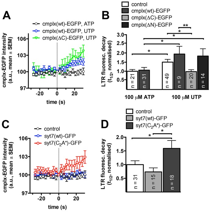
FACE and Ca2+ binding to the C2A domain of Syt7 antagonise complexin-2 recruitment to fused lamellar bodies and thereby facilitate fusion pore expansion. (A) Mean fluorescence of cmplx(wt)–GFP and cmplx(ΔC)–GFP before and after lamellar body fusion with the plasma membrane in cells stimulated with either 100 µM ATP (FACE) or 100 µM UTP (no FACE). FACE inhibits increase of cmplx(wt)–GFP at fused lamellar bodies. Fluorescence change was measured in a peri-vesicular region of interest surrounding individual lamellar bodies. The dotted line indicates the time of fusion (data represent a minimum of ten fusions for each condition). (B) Halftimes of LTR fluorescence decay are not significantly different in cells overexpressing cmplx(wt)–GFP when stimulated with 100 µM ATP (FACE). Stimulation with 100 µM UTP (no FACE) resulted in a significant increase in halftimes of LTR fluorescence decay, which was moderately enhanced in cells overexpressing cmplx(wt)–GFP or cmplx(ΔN)–GFP. In contrast, overexpression of cmplx(ΔC)–GFP completely abolished the increase in the halftime of LTR fluorescence decay following stimulation with 100 µM UTP. (C) Mean fluorescence of cmplx(wt)–GFP before and after lamellar body fusion with the plasma membrane in cells stimulated with 100 µM ATP (FACE). Translocation of cmplx(wt)–GFP to fused lamellar bodies is inhibited in wild-type cells (control, black) and cells expressing Syt7(wt)–GFP (blue) but not in cells expressing Syt7(C2A*)–GFP (red). The dotted line indicates the time of fusion (data represent a minimum of ten fusions for each condition). (D) Halftimes of LTR fluorescence decay in cells overexpressing cmplx(wt)-GFP following stimulation with 100 µM ATP. Deletion of Ca2+-binding to the C2A domain of Syt7 [Syt7(C2A*)–GFP] significantly increased the halftimes of LTR fluorescence decay when compared to wild-type cells and cells expressing Syt7(wt)–GFP. Results are mean±s.e.m. *P<0.05.