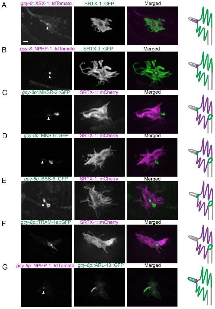
Fig. 2.
Localization of ciliary and dendritic protein markers uncovers the AFD finger compartment as a cilium-related subcompartment. Fluorescently tagged ciliary proteins were produced specifically in the AFD neurons and co-expressed with SRTX-1, a marker of the AFD finger membrane. The IFT-dynein subunit XBX-1 (A) and transition zone protein NPHP-1 (B) are enriched at the base of the cilium. The transition zone proteins MKSR-2 (C) and MKS-6 (D) are localized at the base of the cilium and are also present as ring structures between the finger and dendritic membranes. (E) BBS-8 is also localized in the cilium and at the ring structure. (F) TRAM-1a, which is normally found at the dendritic tips but not inside cilia, is present at the ring outside of the finger compartment in AFD neurons. (G) Co-expression with ARL-13 confirmed that NPHP-1 signal is at the base of the cilium. Arrowheads indicate the base of the cilium. Scale bar: 1 µm.

