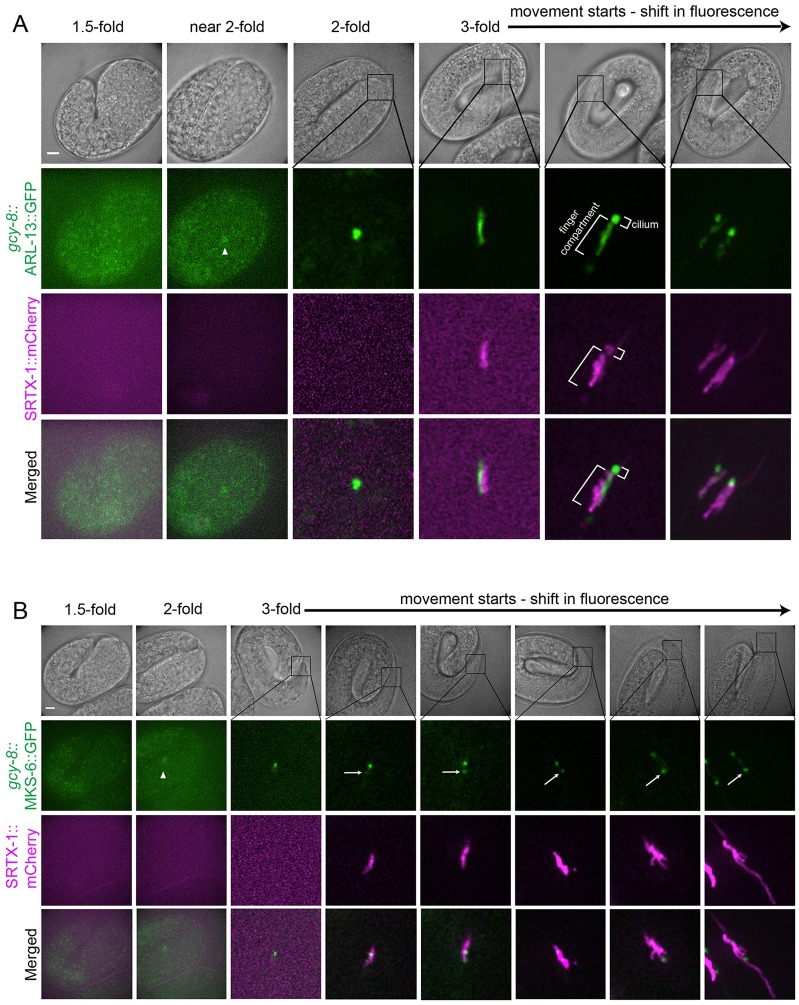
Fig. 3.
Development of the cilium precedes finger formation. The development of the ciliary and finger compartments in AFD neurons are shown at various stages during embryogenesis. Non-overlapping signals at the 3-fold stage are caused by slight movement of the embryos, which could not be completely eliminated without affecting their development during the experimental time period. (A) Ciliary membrane (marked by ARL-13) appears before the fingers (marked by SRTX-1) are formed. (B) The transition zone protein MKS-6 appears first at the base of the cilium, and then at a more posterior second location (arrows), as the finger compartment (marked by SRTX-1) develops. Arrowheads indicate AFD cell bodies. Scale bars: 5 µm.

