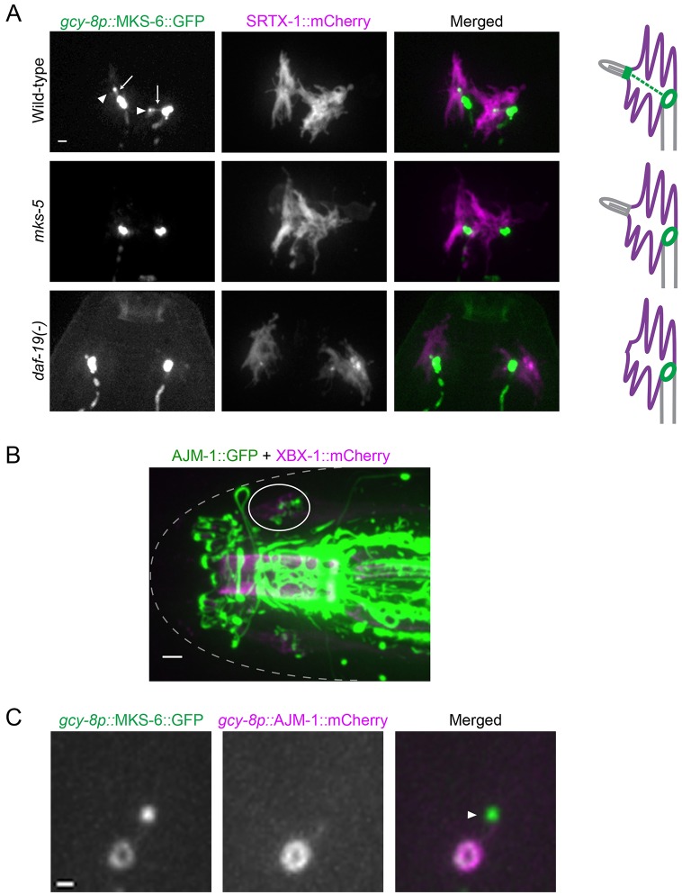
Fig. 4.
An apical junction separates the finger compartment from the dendritic membrane. (A) The ring-like structure is still present in mutants lacking the ciliary transcription factor DAF-19 or the essential transition zone protein MKS-5. Two neurons are shown for each worm. GFP signals are overexposed to highlight the complete absence of the canonical transition zone in the mutants (arrowheads in wild-type). Overexposure also shows a filamentous structure in wild-type worms decorated with MKS-6 that could be IFs (arrows). Scale bar: 1 µm. (B) In the head, apical junctions (marked by AJM-1) are seen as ring-like structures at the base of cilia (marked by XBX-1) in the bilateral amphid channels (one side is circled). Scale bar: 2 µm. (C) AJM-1 is colocalized with MKS-6 at the ring in AFD neurons. Note the absence of AJM-1 at the canonical transition zone (arrowhead). Scale bar: 0.5 µm.

