Fig. 5.
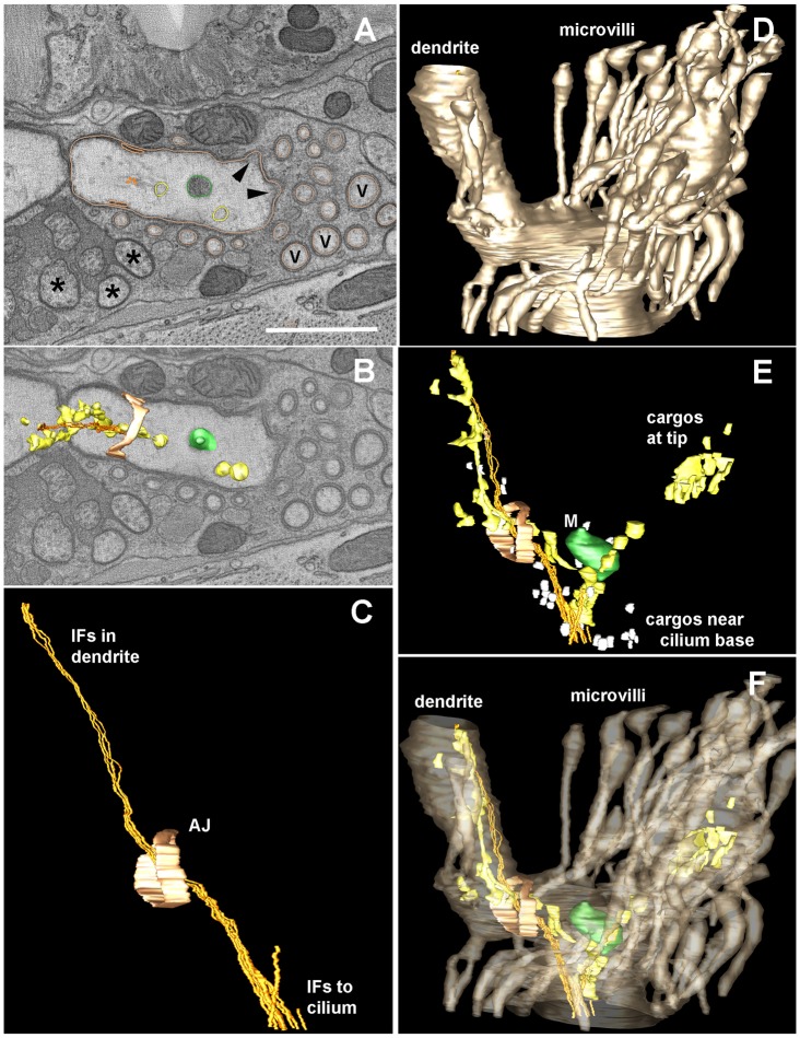
Electron tomography reveals the structure of the AFD dendritic ending. An electron tomogram was produced from thick serial sections for a distal portion of the AFD dendrite, in order to highlight details of the microvilli, the apical junction (AJ, pale orange) to the surrounding amphid sheath cell, and the various contents within the finger compartment, which lie along a cluster of IFs that span from the distal dendrite to the base of the cilium. (A) Orthoslice through a portion of the tomogram showing a variety of objects that have been modeled using IMOD to trace their contours, including a mitochondrion (green) and larger vesicles (beige) that might represent smooth ER. Several closely spaced IFs (gold) run as a tight bundle, with these other small objects lying close by. No microtubules were identified within this region. Several amphid channel cilia (asterisks) and several additional microvilli (V) that were not traced are indicated. Arrowheads indicate the base of two traced microvilli; their cytoplasm is locally more electron dense, representing diffuse actin just beneath the plasma membrane. The outline of traced microvilli is shown in the same color as the AFD plasma membrane (beige). Scale bar: 1 µm. (B) A nearby orthoslice shows three-dimensional features of several modeled objects to better display how they fit within the distal dendrite. (C) Side view of modeled region highlights the extension of the IF bundle from inside the distal dendrite, running past the apical junction to enter the finger compartment. This IF bundle continues anteriorward, outside of the tomogram to reach the base of the cilium (not shown), further anterior to the tomogram. (D) Lateral view of the modeled region highlighting the plasma membrane of AFD (in beige), including the distal dendrite and part of the finger compartment. (E) Same model view as C and D, showing the multiple cargoes that lie along the IF bundle. Close to the base of the cilium, many small vesicles (white) cluster near the IF bundle. Smooth ER clusters along the IF bundle both inside the dendrite and within the finger compartment. (F) Same view as panels C, D, E, highlighting the clustering of cargos inside the finger compartment, but with no cargos entering into the surrounding microvilli.

