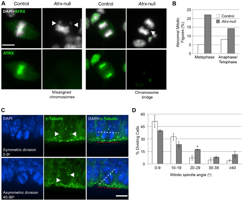
Fig. 1. Atrx-null NPCs exhibit mitotic defects in vitro and altered cell division axis in vivo.
(A) ATRX immunostaining in NPC cultures. DAPI was used as a counterstain. Arrowheads indicate abnormal mitotic figures. Scale bar: 10 µm. (B) Mitotic cells were scored for presence of misaligned chromosomes at the metaphase plate and chromosome bridges or lagging chromosomes at anaphase/telophase (control n = 161; Atrx-null n = 114, from 3–4 embryos). (C) γ-Tubulin immunostaining in E13.5 control and Atrx-null cortex. Arrowheads indicate the centrosomes. The axis of NPC division at the neuroepithelial surface was measured using the axis between centrosomes (white dashed line) relative to the neuroepithelial surface (red dashed line). Scale bar: 10 µm. (D) Angle of division was scored in control and Atrx-null cortex (n = 150 cells; 3 pairs). Data expressed as mean ± S.E.M. *P<0.05.

