
Fig. 6. anti-GFP-DARPin fusion proteins can relocalize fluorescent fusion proteins in D. rerio embryos.
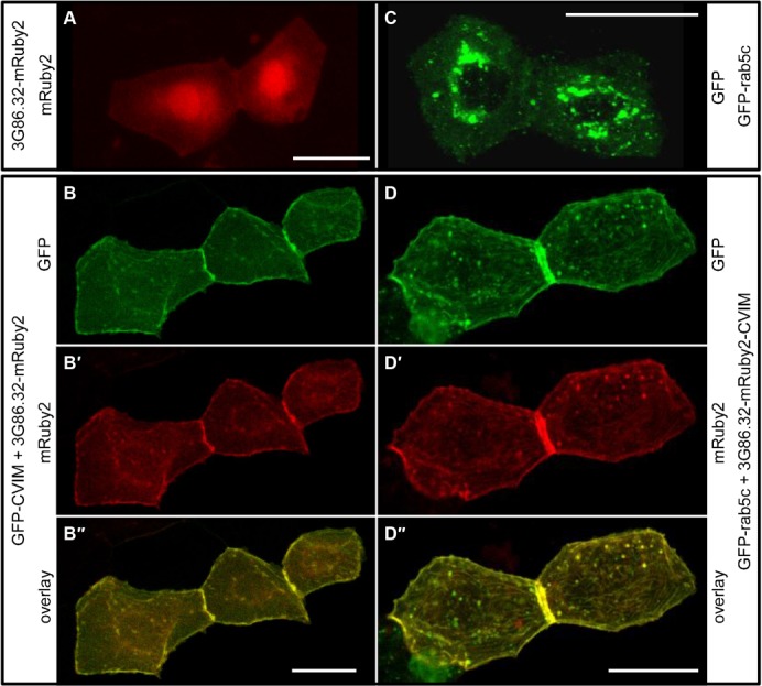
(A,B) 3G86.2-mRuby2 binds GFP in living zebrafish embryos. (A) Control embryo showing the localization of 3G86.32-mRuby2 in two adjacent skin cells. (B–B″) In a zebrafish embryo co-expressing membrane-bound GFP-CVIM (B), 3G86.32-mRuby2 now localizes to the plasma membrane (B′) and shows a virtually complete co-localization with GFP-CVIM (B″). (C,D) membrane-anchored 3G86.32-mRuby2-CVIM recruits GFP-rab5c in living zebrafish embryos. (C) Control embryo showing the localization of GFP-rab5c in two adjacent skin cells. (D–D″) In a zebrafish embryo co-expressing membrane-bound 3G86.32-mRuby2-CVIM (D′), GFP-Rab5C also localizes to the plasma membrane (D) and shows a virtually complete co-localization with 3G86.32-mRuby2-CVIM (D″). Scale bars are 20 µm.