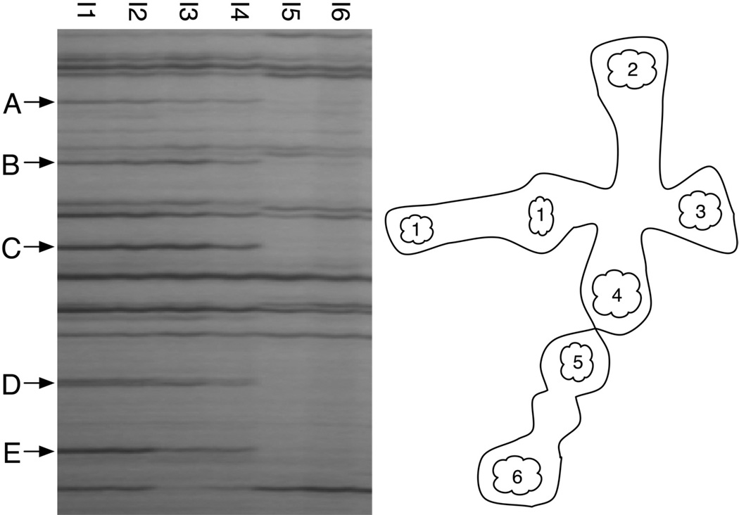
Figure 4.
AFLP fingerprints of somatic samples from bichimera 4497-7 show the two distinct genotypes are spatially segregated within the colony. Each lane of the gel corresponds to a somatic sample of pooled intestines (I1–I6); location of each sampling site in the chimera is illustrated on the right. Note the repeatability of independent samples I1-I4 (arrowheads outline unique presence bands in these samples), and I5 and I6, which are also equivalent.

