
Figure 3.
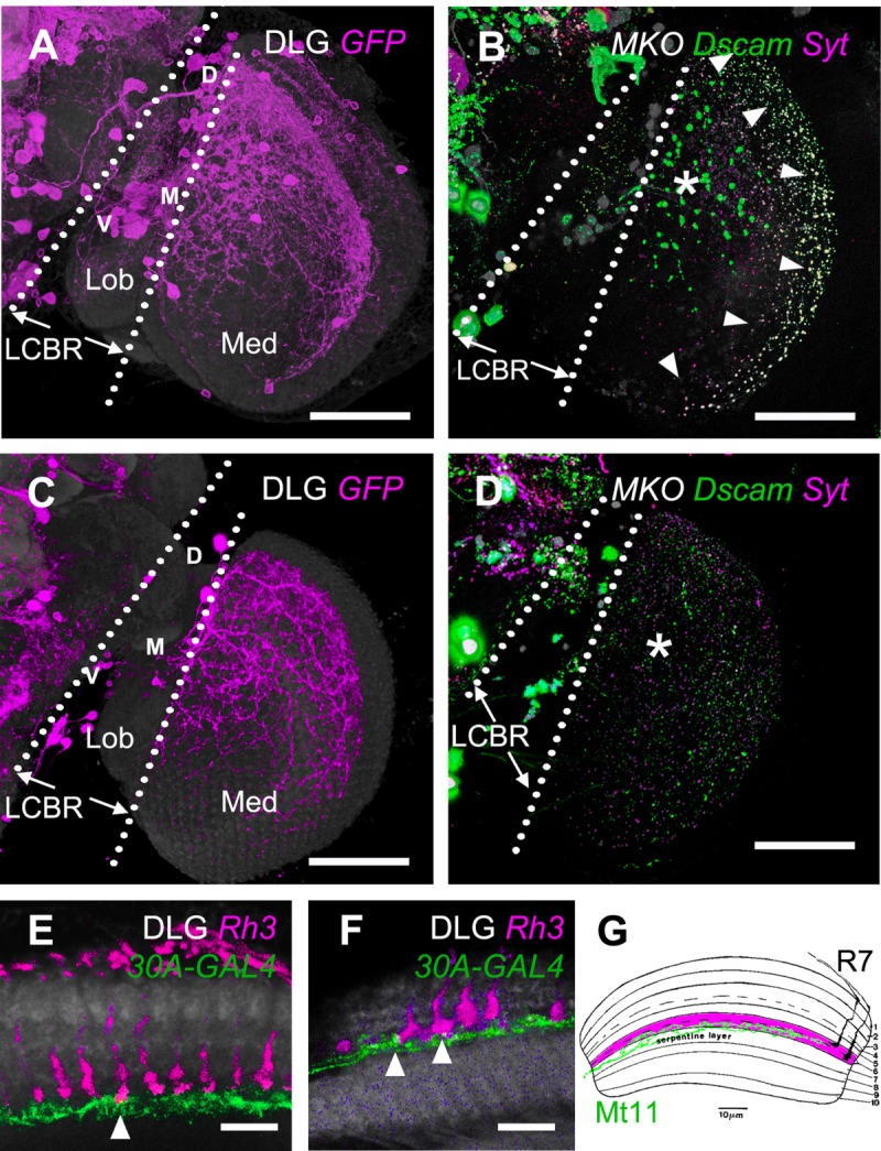
A specific driver of M6-LNs. A: Expression pattern of 30A-GAL4 (magenta). The brain is immunostained with anti-DLG (gray). Locations of M6-LN cell bodies at the ventral (V), medial (M), and dorsal (D) LCBR are marked. B: Presynaptic marker Syt::HA (magenta) and postsynaptic marker Dscam-GFP (green) in the 30A-GAL4 neurons labeled by MKO (gray). C: Expression pattern of 30A-GAL4;Cha-GAL80. The brain is immunostained with anti-DLG (gray). D: Presynaptic marker Syt::HA (magenta) and postsynaptic marker Dscam-GFP (green) in the 30A-GAL4;Cha-GAL80 neurons labeled by MKO (gray). The intense Dscam-GFP signal at the anterior M6 (asterisk), and the overlapping Dscam-GFP/Syt::HA signal at the posterior M6 (arrowhead) in B is absent in D. LCBR, lateral cell body rind; Med, medulla; Lob, lobula. E,F: Dual expression of 30A-GAL4 (green) and Rh3-LexA (magenta) in anti-DLG immunostained medulla (gray). At the medial M6 (E), only long-form R7 (arrowhead) intersects with M6-LNs (green). At the dorsal-posterior M6 (F), all R7s intersect with M6-LNs. G: A merged illustration of Mt11 (green) and two forms of R7 in medulla from Fischbach and Dittrich (1989). The restriction of the terminals of Mt11 to the proximal half of layer M6 is similar to that of the M6-LNs expressed by 30A-GAL4. Scale bars = 50 μm in A–D; 10 μm in E,F.