
Figure 9.
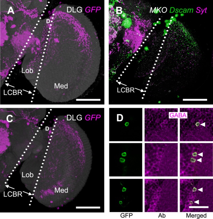
M6-LNs in VT12760-GAL4. A: Expression pattern of VT12760-GAL4 (magenta). M6-LNs cell bodies are located at the dorsal (D) LCBR. The brain is immunostained with anti-DLG (gray). B: Polarity labeling with presynaptic marker Syt::HA (magenta) and postsynaptic marker Dscam-GFP (green) in the M6-LNs labeled by MKO (gray). C: VT12760-GAL4 expression pattern subjected to Cha-GAL80 inhibition (magenta). The brain is immunostained with anti-DLG (gray). D: Remaining cell bodies of M6-LNs in VT12760-GAL4 after Cha-GAL80 inhibition (green) are all anti-GABA (magenta) immunopositive (arrowheads). LCBR, lateral cell body rind; Med, medulla; Lob, lobula. Scale bars = 50 μm in A–C; 10 μm in D.