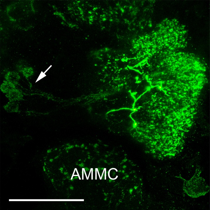
Figure 5.
FMRFamide immunoreactivity in the AL of a female Ae. aegypti. Maximum projection of 37 optical sections showing a frontal image of an AL, where FMRFamide immunoreactivity is observed in all AL glomeruli. In addition, thick varicose FMRFamide-immunoreactive fibers of an extrinsic neuron are visible at the center of the AL neuropil. The arrow indicates the FMRFamide-immunoreactive cell bodies in the lateral cell cluster. AMMC: antennal motor and mechanosensory center. Scale bar = 25 μm.

