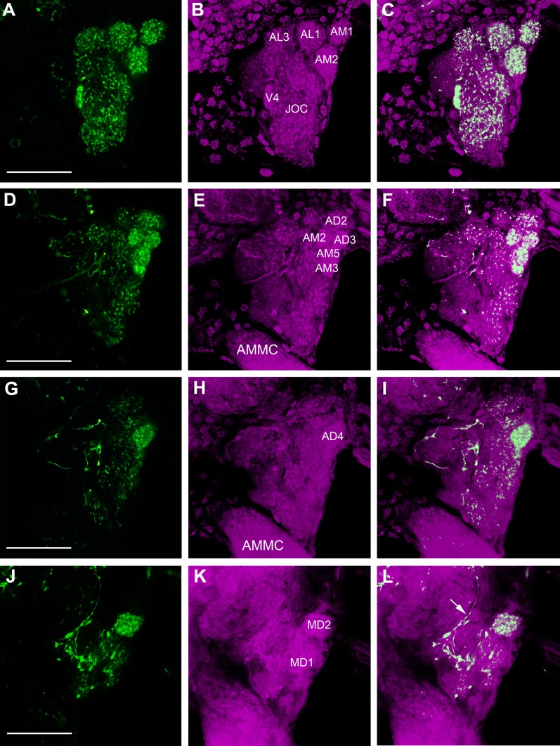
Figure 6.
Detailed innervation pattern of FMRFamide-immunoreactive fibers (green) in the AL neuropil (magenta) of a female Ae. aegypti. A–C: Maximum projection of six optical sections showing a frontal image of the AL, where strong FMRFamide-immunoreactivity is observed in the anteromedial (AM1, AM2) and anterolateral glomeruli (AL1, AL3), as well as in a ventral glomerulus (V4). Note the compartmentalization of immunoreactivity in the AL glomeruli and the Johnston’s organ center (JOC). D–I: Maximum projections of six optical sections showing the strong immunoreactivity in the anteromedial (AM2, AM3, AM5) and anterodorsal glomeruli (AD3, AD4). Note that the lateral part of the AL is almost devoid of immunoreactivity. Coarse varicose fibers are also seen in this posterolateral region of the AL. J–L: Maximum projection of five optical sections showing the axon (arrow) of the FMRFamide-immunoreactive extrinsic neuron that connects the protocerebrum and the AL. The varicose fibers of this neuron converge on the posterior and lateral side of the AL. At this level the varicose neuron starts to wrap around the maxillary palp glomerulus, MD1, sparing neighboring glomeruli like MD2. AMMC: antennal motor and mechanosensory center. Scale bars = 25 μm.

