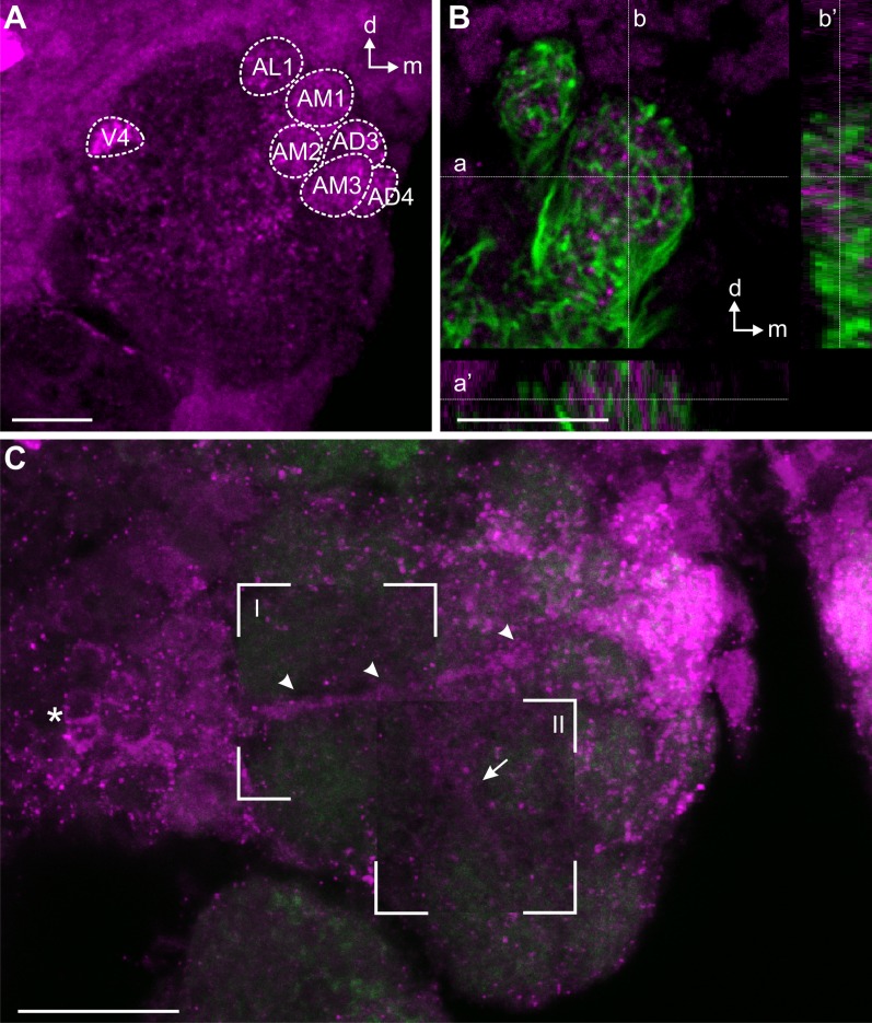
Figure 8.
sNPF immunoreactivity (magenta) in the AL of a female Ae. aegypti. A: Maximum projection of 38 optical sections, showing six strongly labeled glomeruli in the anterior part of the AL (AL1, AM1–3, AD3, AD4) and one strongly labeled glomerulus in the ventral area of the AL (V4). B: Comparison of sNPF immunostaining (magenta) with back-filled OSN fibers (green) revealed no overlap between sNPF and OSN profiles. Confocal image stack of 13 optical images. Insets below and to the right show the z-stack along lines a and b, whereas lines a′ and b′ mark the position of the main optical image within the stack. C: Collage of maximum projections containing different numbers of optical sections from the same stack of 16 optical sections, revealing a cell cluster lateral to the AL sending axons to the AL (arrowheads) innervating the glomeruli. The background maximum projection contains sections 1 to 7, and maximum projections shown in I and II contain optical sections 7 to 16 and 11 to 13, respectively. Green, synapsin immunostaining; compare with FMRFamide immunostaining in Figs. 5, 6. A–C: Z-distances between optical sections: 0.5 μm. Scale bars = 10 μm.

