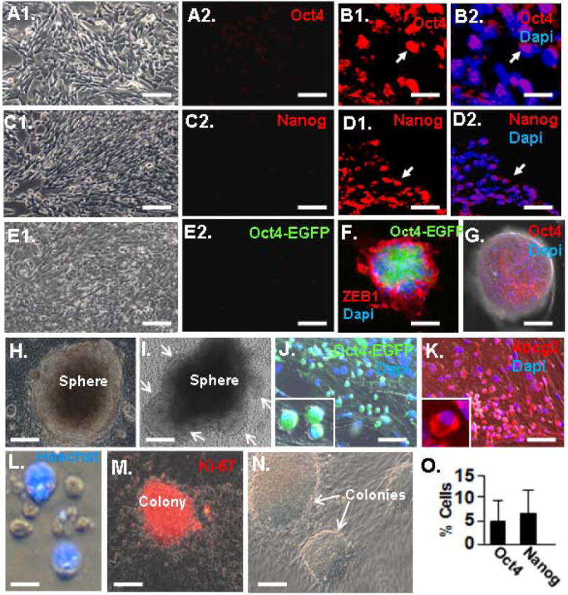
Figure 2.

Sphere-induced induction of Oct4 and Nanog. A. Immunostaining for Oct4 in MEF monolayer culture. A1 is a phase image. B. Three-week-old spheres were sectioned and immunostained for Oct4. Note nuclear Oct4 expression. Arrows indicate the same position in the panels. C. Monolayer cultures of MEFs immunostained for Nanog. C1 is a phase image. D. Immunostaining of sphere section for Nanog as in panel B. Note nuclear Nanog expression. E. No EGFP expression is evident in monolayer cultures of tail tip fibroblast isolated at birth from Oct4-EGFP mice. E1 is the phase image. F. Sphere fromed from Oct4-EGFP fibroblast after two weeks in suspension culture immunostained for Zeb1 and showing EGFP immunofluorescence. G. Similar sphere to that in panel G immunostained for Oct4. H. Three-week-old sphere bound to a Matrigel-coated plate. I. Sphere similar to that in panel H, three days after adhesion. Arrows show cell migrating out of the sphere. J. Small, round and poorly adherent Oct4-EGFP+ cells evident at the site of sphere dissociation, one week after sphere adhesion to Matrigel. K. Similar cells to those in panel J immunostained for Abcg2. L. Cell rinsed from plates in panels K and J are enriched in a population of small, Hoechst- cells. M. Cells from panel L from colonies that immunostain for the proliferation marker Ki-67. N. Phase image of colonies. O. Quantification of Oct4+ and Nanog+ cells in three-week-old spheres. The bar is 10 μm in panels B and D, 15 μm in panel L, 20 μm in panels A, C, E, J, K, M an N, and 100 μm in panels F and G.
