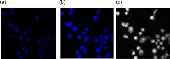
Figure 5.

Fluorescence images of MDA-MB-231 cells incubated with 50 μM complex 1 for 2 h at 37 °C (λex, 350 nm; λem, 390 nm; short pass DAPI filter cube). (a) First image without light exposure, (b) image recorded after three 10 s pulses of low power LED light, and (c) gray scale version of image (b).
