
Fig. 1.
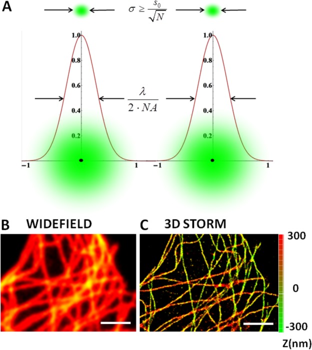
Direct 3D STORM images of microtubule network in Hela cells. Panel A illustrates the principle of localization precision (σ) which replaces diffraction limit by precisely points out the single molecule position by a Gaussian fit over a number of frames (N). In panels B and C, α-tubulin is immunostained with Alexa 647 anti-mouse secondary antibody in order to perform direct STORM imaging. The comparison between the conventional wide field image (1B) and the superresolution image obtained using 3D STORM (1C).The axial position is represented as a Z-color coded map and the scale bar is 2 µm. Image was reconstructed from 1000 frames of 20 ms exposure time to reach a localization precision ∼20 nm along the radial direction and ∼60 nm along the axial one.