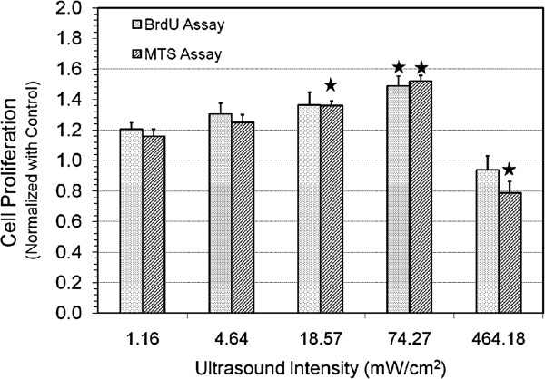
Figure 2.

Change in normalized proliferation of MC3T3-E1 cells with ultrasound intensity. Change in proliferation of ultrasound stimulated MC3T3-E1 cells (normalized with control) at different ultrasound intensities (SATA) with frequency = 1.5 MHz, PRF = 1 kHz, pulse duration = 200 μs and exposure time = 10 min. Values significantly different from control group have been indicated by filled stars for p < 0.05.
