
Figure 4.
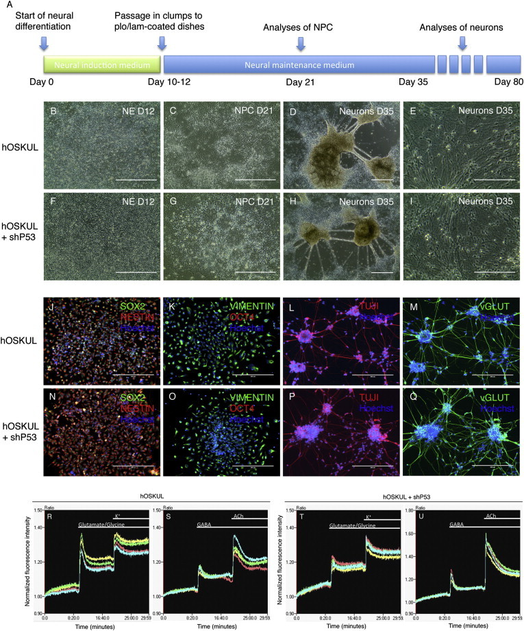
iPSCs Generated by Transient p53 Suppression Can Differentiate to Functional Neurons In Vitro
(A) Timeline showing the directed neural differentiation of induced pluripotent stem cell (iPSC) lines generated with or without (w/wo) a short hairpin to p53 (shp53).
(B–I) Phase contrast morphology of neuroepithelium (NE) at day 12 (B and F), neural progenitor cells (NPC) at day 21 (C and G), and neurons at day 35 (D, E, H, and I).
(J–Q) Immunocytochemistry of NPCs at day 21 with SOX2, NESTIN, and Hoechst (J and N) and VIMENTIN, OCT4, and Hoechst (K and O) and of neurons at day 35 with TUJI and Hoechst (L–P) and vGLUT and Hoechst (M and Q).
(R–U) Intracellular calcium kinetics in iPSC-derived neurons generated without (R and S) or with shp53 (T and U). Baseline fluorescence was recorded for 10 min before application of 300 μM glutamate/10 μM glycine, 25 mM K+, 100 μM GABA and 300 μM acetylcholine. The fluorescence was normalized to the first data point of each of the traces.
Scale bars, 200 μm (B–D, F–H, and J–Q) and 100 μm (E and I).