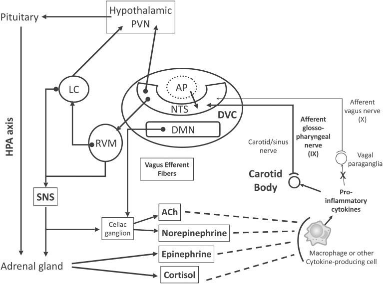
Figure 1.
Proposed model for inflammation-induced central nervous system (CNS) activation through carotid body (CB) chemoreceptors and subsequent efferent neuroimmunomodulator activation. Briefly, chemosensory transduction begins in immune cells, which release inflammatory mediators (i.e., TNF-α, IL-1β, and IL-6) to activate the CB. The CB is innervated by glossopharyngeal (cranial nerve IX) afferent neurons, the cell bodies of which are located in the petrosal ganglion, and their central projections end primarily within the dorsal vagal complex (DVC) of the medulla oblongata. The DVC consists of the nucleus tractus solitarii (NTS), the dorsal motor nucleus of vagus nerve (DMN), and the area postrema (AP). The DMN is the main site of origin of preganglionic vagus efferent fibers, which evoke acetylcholine (ACh) release from celiac ganglion neurons, inhibiting the pro-inflammatory response. The AP lacks a brain-blood barrier (dotted line, AP), projects into the NTS and is an important site for humoral immune-to-brain communication. The main portion of the CB chemosensory inputs is received by neurons in the NTS, which coordinate autonomic function and interaction with the endocrine system. Ascending projections from the NTS reach the hypothalamic paraventricular nucleus (PVN), an important structure involved in hypothalamus-pituitary-adrenal (HPA) axis activation, releasing cortisol, which, in fact, inhibits the pro-inflammatory response. Synaptic contacts also exist between neurons in the NTS and rostral ventrolateral medulla (RVM), which plays an important role in the control of cardiovascular and respiratory homeostasis. Neurons from the RVM project to the locus coeruleus (LC), which innervates higher brain sites, such as the PVN. Neuronal projections emanate from the RVM and LC to sympathetic preganglionic neurons (SNS) in the spinal cord, which innervates both the adrenal medulla and celiac ganglion for epinephrine and norepinephrine release, respectively, inhibiting the pro-inflammatory response. These ascending and descending connections provide a neuronal substrate for neuroimmunomodulatory mechanisms. Excitatory pathways, continuous lines; inhibitory pathways, dashed lines.

