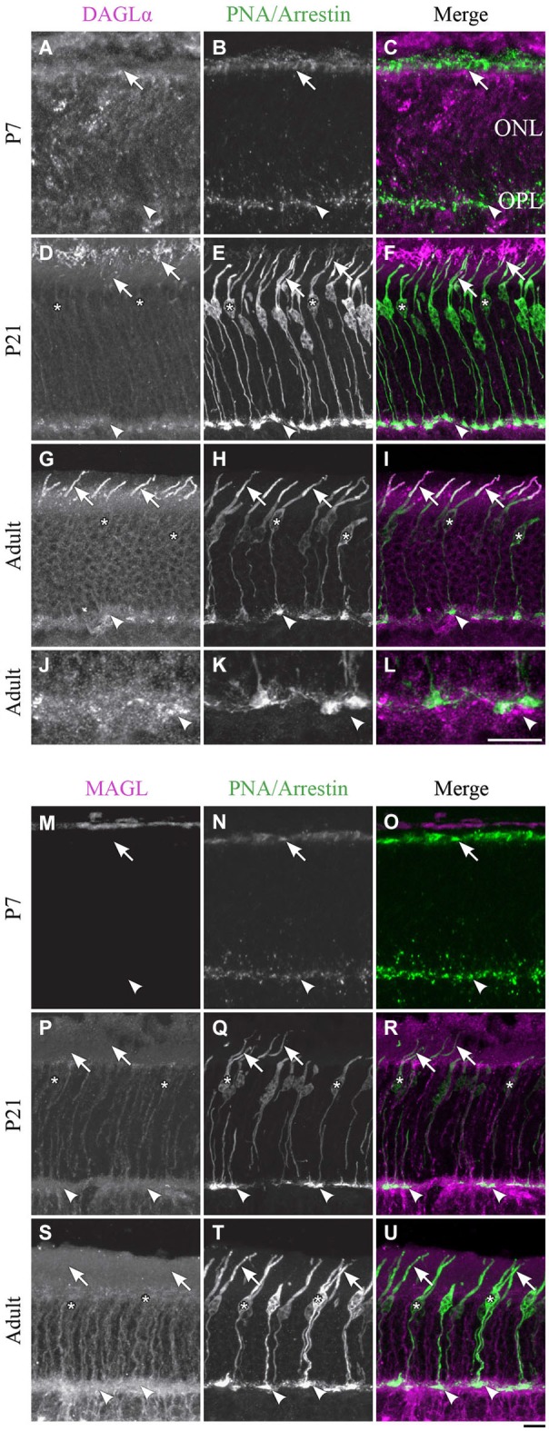
Figure 3.

DAGLα and MAGL immunoreactivity in cone photoreceptors. (A–U) confocal micrographs of P7, P21 and adult rat retinas co-labeled for DAGLα (A–L) or MAGL (M–U) and the cell-type specific marker for cone photoreceptors, PNA (for P7) or cone-arrestin (for P21 and adult). Each protein is presented alone in gray scale: DAGLα or MAGL in the first column and PNA/cone-arrestin in the second; then the two are presented merged in the third column (DAGLα or MAGL in magenta and PNA/cone-arrestin in green). DAGLα is localized in the outer (arrows) and inner segments of cones, as well as the cell body (stars) but not the synaptic pedicle (arrowheads). MAGL is not detectable in any part of the cone photoreceptors. ONL, outer nuclear layer; OPL, outer plexiform layer. Scale bar = 10 µm.
