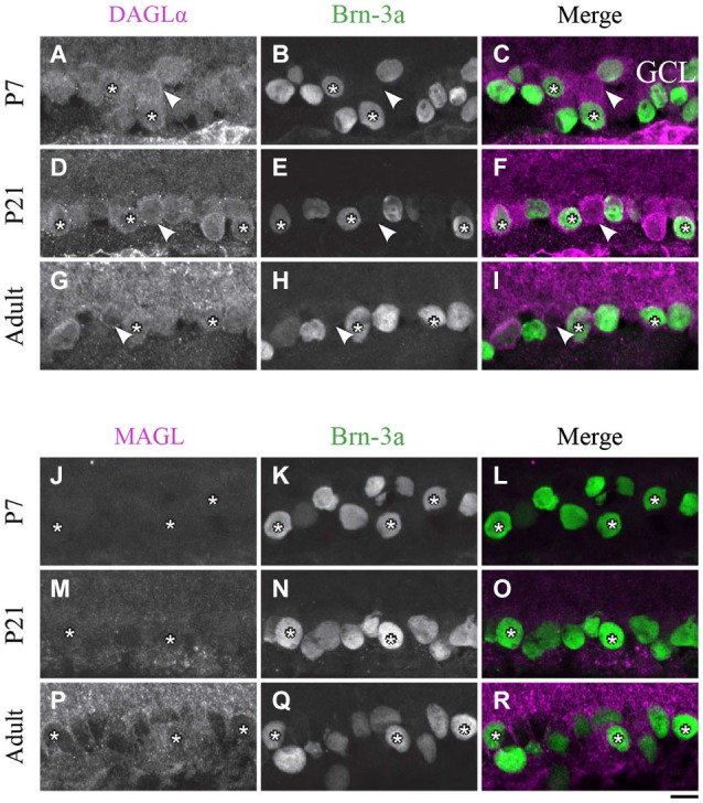
Figure 9.

DAGLα and MAGL immunoreactivity in ganglion cells. (A–R) confocal micrographs of P7, P21 and adult rat retinas co-immunolabeled for DAGLα (A–I) or MAGL (J–R) and the cell-type specific marker for the ganglion cells, Brn-3a. DAGLα is localized in the cell bodies (stars) of the ganglion cells as well as in displaced amacrine cells or intrinsically photosensitive retinal ganglion cells (ipRGCs) (arrowheads) from P1 to the adult age. MAGL is not detectable in the ganglion cells. GCL, ganglion cell layer. Scale bar = 10 µm.
