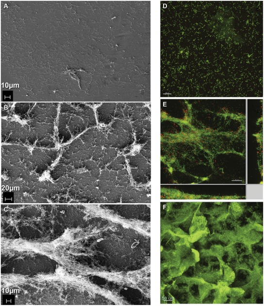
FIGURE 3.

Formation of coculture biofilm over time. (A–C) Electron micrographs of fixed coculture at (A) early (386X, 0 h; B) intermediate (243X, 48 h; C) steady-state (336X, 240 h) time points. (D–F) Fluorescence micrographs of coculture biofilm (D,E) embedded in polyacrylamide and hybridized with domain-specific oligonucleotide probes labeled green for D. vulgaris and red for M. maripaludis at (D) early (0 h) and (E) intermediate (48 h) time points. (F) Intact hydrated biofilm unfixed and stained with Acridine Orange at steady-state (240 h).
