
Figure 2.
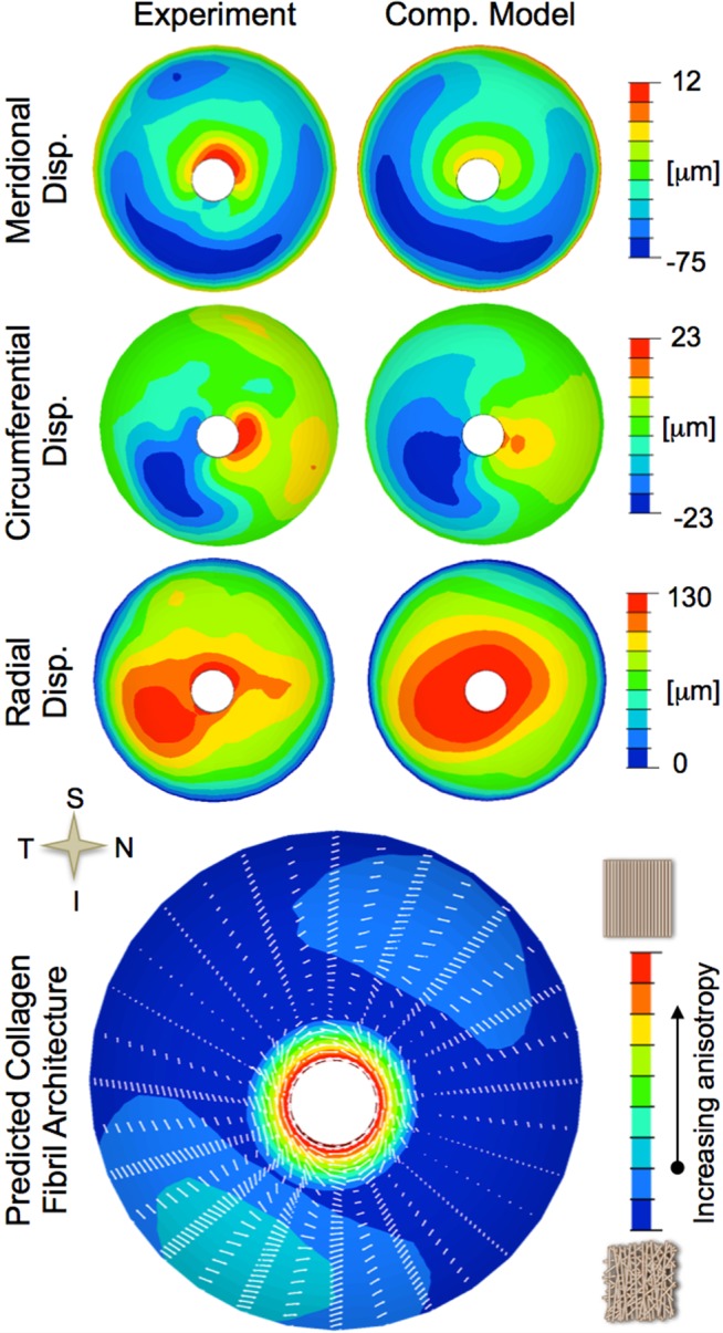
Typical inverse numerical characterization of a scleral shell (left eye of a 77-year-old donor of African descent). Top: Comparison of experimentally measured displacements and the displacements predicted by the inverse computational model for an IOP elevation from 5 to 45 mm Hg. Bottom: Predicted anisotropic collagen architecture showing the predominant collagen fibril orientations (white lines) and the degree of anisotropy (contour plot). A distinct ring of circumferentially-aligned fibrils can be seen around the scleral canal, which was common to all eyes.