
Figure 3.
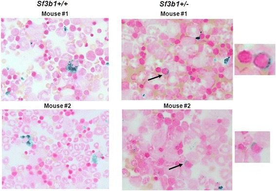
Detection of ring sideroblasts by Prussian blue staining in Sf3b1 +/− compared to Sf3b1 +/+ mice. Bone marrow cells were extracted from femurs of Sf3b1 +/− (n = 5) and Sf3b1 +/+ (n = 5) and cells (2-3x10⁵) spotted on cytospin slides prior staining with Prussian blue. Ring sideroblasts (RS) were detected in Sf3b1 +/− compared to Sf3b1 +/+ mice. Images were taken from 2 mice per group. RS were also detected in additional mice as shown in Additional file 5: Figure S5.