
Figure 4.
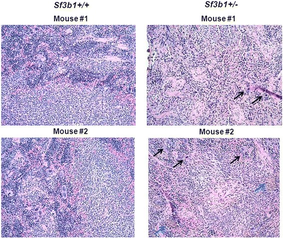
Histology of splenic tissues of Sf3b1 +/− compared to Sf3b1 +/+ mice. Spleens from Sf3B1 +/− and Sf3b1 +/+ mice were fixed in 4% formaldehyde/PBS and embedded in paraffin. Sections were stained with Haematoxylin–Eosin and showed extramedullary hematopoiesis with all 3 hematopoietic elements, increased megakaryocytes with hyperchromatic nuclei (black arrows), increased hemosiderin deposits (blue arrows) and evidence of fibrosis.