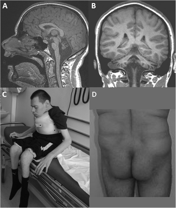
Figure 1.

Brain MRI and morphological aspects of adult PMM2-CDG. Upper panel. Brain MRI of patient #19 at 16 years old. T1-weighted sagittal (A) and frontal (B) images showing severe atrophy of the cerebellum, including vermis and hemispheres, and less atrophied pons. Lower panel. Picture of a 32 year-old patient with severe ataxia, neuropathy and severe intellectual disability (C). Picture of a 45 year-old patient showing the abnormal fat repartition typical of PMM2-CDG (D).
