
Figure 6.
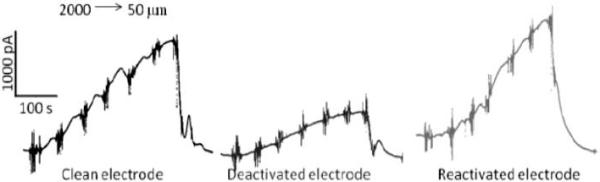
Oxidation current versus the diamond microelectrode-mucosa spacing. The measurements were made by continuous amperometry in the mouse ileum at 0.8 V vs. Ag/AgCl. The tissue was mounted in a bath with perfusing Krebs buffer (pH 7.4). Approach curves were recorded from 2000 to 10 μm for a clean electrode, after the electrode was deactivated and after reactivation by galvanostatic pretreatment. Error bars represent standard deviations for three approach curves measured for each electrode in the same tissue.