
Figure 1.
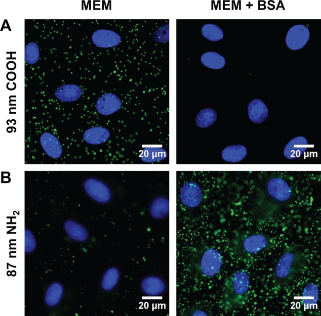
Fluorescence microscopy images show cellular binding of NPs (green) in MEM and MEM supplemented with 10 mg mL–1 BSA (MEM + BSA) to monkey kidney epithelial cells (BS-C-1) at 4 °C. (A) 93 nm carboxylate-modified NPs. (B) 87 nm amine-modified NPs. Nuclei are stained with DAPI (blue).