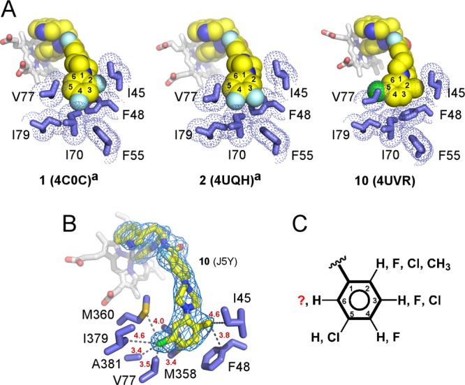
Figure 6.

Buried binding mode. (A) van der Waals interactions between the terminal N-phenyl rings of 1, 2, and 10 (yellow spheres) and amino acid side chains (blue sticks). PDB codes of the corresponding structures are shown in parentheses. (B) The fragment of the 2fo – fc electron density map (blue mesh) contoured at 1σ demonstrates unambiguous orientation of the terminal ring placing the 5-chloro substituent at van der Waals distances of V77, M358, M360, I379, and A381. Heteroatoms are colored by types: oxygen in red, nitrogen in blue, fluorine in cyan, chlorine in green. Heme is shown in gray sticks. Distances in red are in angstroms. (C) Collective substitution pattern of the terminal N-phenyl ring deduced from the X-ray structure analysis and SAR. Superscript “a” indicates that cocrystal structures with compounds 1 and 2 were previously published.13
