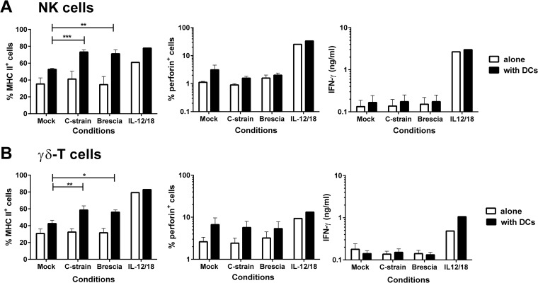
FIG 1.
Analysis of the direct and indirect effects of CSFV on NK and γδ T cells. Enriched NK and γδ T cells were cocultured with an attenuated C-strain or virulent Brescia strain of CSFV, either alone or with enriched blood DCs, at a ratio of 2:1. A mock virus-infected cryolysate supernatant and a cocktail of recombinant porcine IL-12 and IL-18 were used as negative and positive controls, respectively. Twenty-four hours postculture, the percentages of MHC-II- and perforin-expressing cells were assessed on the NK cells (A) and γδ T cells (B) by flow cytometry, and the IFN-γ content of the culture supernatants was assessed by ELISA. The mean data ± SEM from three independent experiments utilizing different animals are shown. The values for each virus-stimulated condition were compared to the corresponding mock-stimulated control using a one-way ANOVA, followed by Dunnett's multiple comparison test: ***, P < 0.001; **, P < 0.01; *, P < 0.05.

