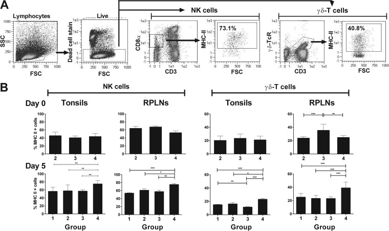
FIG 4.
Ex vivo analysis of the MHC class II expression on lymphoid tissue-derived NK and γδ T cells 5 days following C-strain vaccination and 5 days post-virulent CSFV challenge. The cells were isolated from tonsils and retropharyngeal lymph nodes (RPLN) 5 days after vaccination (day 0) and 5 days after Brescia challenge (day 5), and MHC-II expression on NK and γδ T cells was assessed using flow cytometry. (A) Gating strategy used to interrogate responses in live γδ T cells (CD3+ γδ-TCR+) and NK cells (CD3− CD8αlow). (B) Mean percentage of MHC-II-expressing NK and γδ T cells for each experimental group (group 1, C-strain vaccination [intranasal {i.n.}] and mock challenge; group 2, C-strain vaccination [intramuscular {i.m.}] and CSFV Brescia challenge; group 3, C-strain vaccination [i.n.] and CSFV Brescia challenge; group 4, mock vaccination and CSFV Brescia challenge), tissue, and time point. The data represent the mean of 3 pigs/group/time point ± SEM. Statistical analyses were performed using a one-way ANOVA, followed by Bonferroni's multiple comparison test: ***, P < 0.001; **, P < 0.01; *, P < 0.05.

