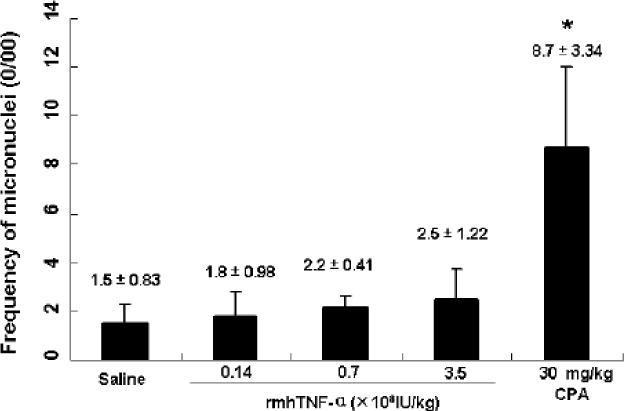
Fig.2.

Frequency of micronuclei in mouse polychromatic erythrocytes after rmhTNF-α treatments (0/00). Activity of rmhTNF-α to induce bone marrow micronucleus was assessed in ICR mice. Three groups (6 mice/group) received 3.5×10⁸, 0.7×10⁸ to 0.14×10⁸ IU/kg i.m. administrations of rmhTNF-α respectively. Saline and cyclophosphamide (CPA, 30 mg/kg) were used as negative and positive controls. 48 hrs later, the animals were sacrificed and the thighbone marrow cells were analyzed following methanol fixing and Giemsa staining. The frequency of micronuclei was counted based on an examination of 1000 polychromatic erythrocytes (PCE) for every mouse. The results did not show any significant difference between rmhTNF-α and saline cohorts (P>0.05) which indicated no micronuclei inducing toxicity for rmhTNF-α in mice.
