
Fig. 3.
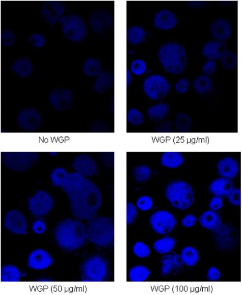
Fluorescence microscopy images of human macrophage monolayers at day 5 of exposure to WGP after staining with a fluorescent cell death marker. MDMs were grown in RPMI containing autologous serum on glass coverslips in a 24-well tissue culture plate for 12 days when the cells were exposed to different concentrations WGP. After washing away β-glucan, the cell monolayers were stained with aqua fluorescent reactive dye, then processed for and analyzed by confocal fluorescence microscopy (Olympus FV1000). Differential intensity of staining was used to count live (low intensity) and dead (high intensity) cells from at least 150 macrophages in each treatment sample in 2 independent experiments. High numbers of cells were scored as dead following treatment with 100 μg/ml WGP.