
Fig. 4.
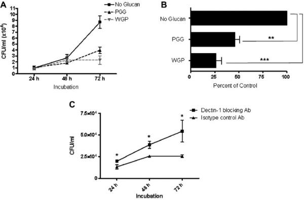
β-glucan inhibits intracellular growth of M. bovis BCG within macrophages through dectin-1. MDMs in each well (2 × 10⁵ cells/well) of a 24-well plate were incubated at 37 °C/5% CO2 in the absence or presence of PGG (50–100 μg/ml) or WGP (25–50 μg/ml). MDMs were then infected with 2 × 10⁵ M bovis BCG (MOI = 1:1) and incubated at 37 °C/5% CO2 for 2 h. The infection was stopped by washing and the cells were incubated in RPMI + 2.0% autologous serum for 24, 48 or 72 h. At each time period, cells were lysed and lysates plated on 7H11 agar for CFU analysis (expressed as CFUs/ml). A) Growth curve of M. bovis BCG showing inhibition of growth at 72 h in both PGG and WGP pre-treated macrophages. B) Results from A expressed in percentage values compared to no-glucan control which is set at 100%. Compared to the control, both PGG and WGP treatment showed a significant decrease in bacterial growth in MDMs by 54 ± 4.8% (**p < 0.005) and 74 ± 5.9% (***p < 0.0005), respectively. C) Growth curve of M. bovis BCG in macrophages pre-treated with anti-Dectin-1 or isotype control antibody, followed by the addition of WGP. Blocking the receptor increased the survival of M. bovis BCG following the addition of WGP. Data shown in A, B and C are from representative experiments (A and B, n = 3; C n = 2; each performed in triplicate).