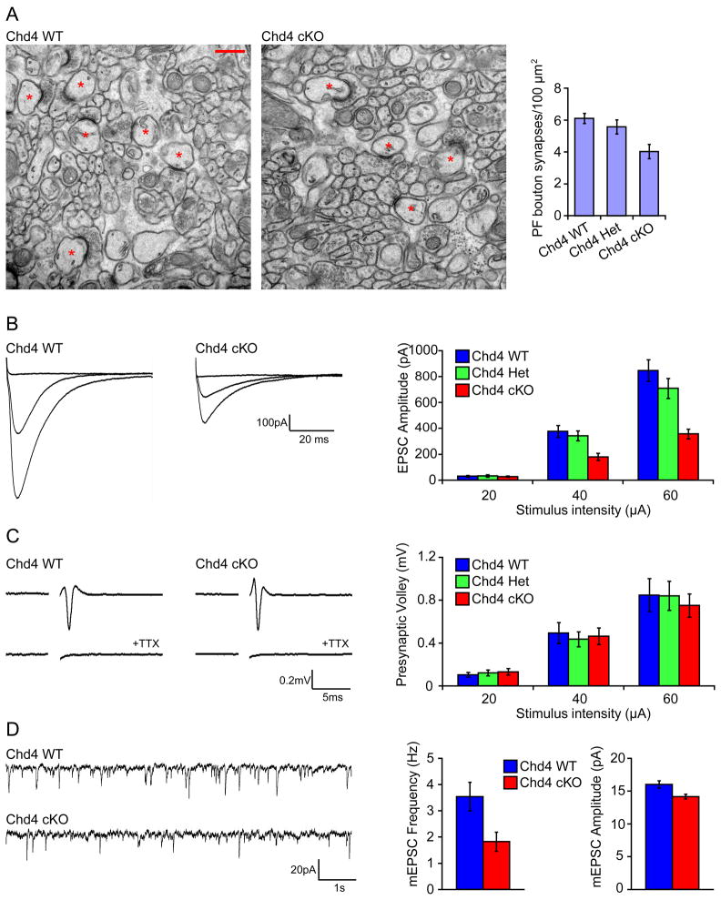
Figure 2. Knockout of the core NuRD complex subunit Chd4 impairs granule neuron/Purkinje cell synaptogenesis and neurotransmission in the cerebellar cortex.
(A) Cerebella from P22 Chd4 conditional knockout mice, Chd4 heterozygous mice, and control Chd4loxP/loxP mice were subjected to electron microscopy analyses. Left panels: representative electron micrographs of the molecular layer of the cerebellar cortex from Chd4 conditional knockout mice (Chd4 cKO) and control Chd4loxP/loxP mice (Chd4 WT) are shown. Synapses comprising of parallel fiber presynaptic boutons apposed to Purkinje cell postsynaptic spines are denoted with asterisks. Scale bar: 500nm. Right panel: quantification of the density of granule parallel fiber/Purkinje cell synapses in Chd4 conditional knockout (Chd4 cKO), Chd4 heterozygous (Chd4 Het), and control Chd4loxP/loxP (Chd4 WT) mice. The density of synapses is reduced in Chd4 conditional knockout mice compared to control Chd4loxP/loxP mice (p<0.005, ANOVA followed by Fisher’s PLSD post hoc test, n=10–12 regions, 2 brains). (B) Acute sagittal cerebellar slices were prepared from P20–P24 Chd4 conditional knockout, Chd4 heterozygous, and Chd4loxP/loxP mice and parallel fiber-evoked Purkinje cell currents (PF-EPSCs) were recorded in response to increasing stimulus intensities (20, 40, and 60μA). Representative current traces (left panels) and quantification of the PF-EPSC amplitude (right panel) are shown. Chd4 conditional knockout mice (Chd4 cKO) have reduced evoked EPSC amplitude compared to control Chd4loxP/loxP mice (Chd4 WT) (p<0.001, ANOVA followed by Fisher’s PLSD post hoc test, n=21–26 neurons, 5 brains). (C) Acute coronal cerebellar slices were prepared as in (B), and parallel fiber axons were stimulated at sites 400 μm away from an extracellular recording electrode. A representative trace of the stimulus-evoked presynaptic waveform before and after application of TTX is shown in the left panels. The stimulus artifact was removed for clarity. Quantification of presynaptic volley amplitude is shown in the right panel. Conditional knockout of Chd4 has little or no effect on the presynaptic volley amplitude. (D) Acute sagittal slices cerebellar were prepared as in (B) and Purkinje cell mEPSCs were recorded in the presence of TTX. Representative traces of mEPSCs from Chd4 conditional knockout and control Chd4loxP/loxP mice are shown in the left panel. Quantification of the mEPSC frequency and amplitude are shown in the right panels. Chd4 conditional knockout mice had reduced mEPSC frequency and amplitude compared to control Chd4loxP/loxP mice (p<0.05, t-test, n=24–27 neurons, 7 brains).

