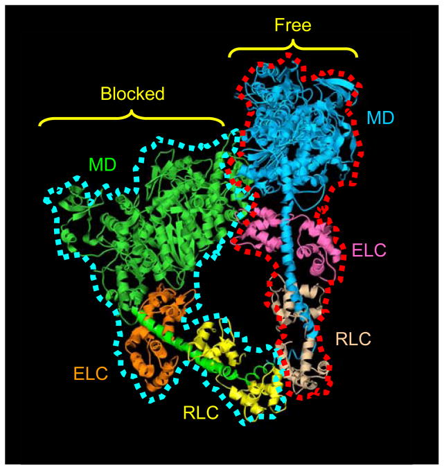
Figure 1. Atomic model of head-head interaction.
This ribbon model represents the best fit of the atomic model of vertebrate smooth muscle HMM (PDB1i84)9 into the tarantula 3D reconstruction.11 The actin-binding region of the blocked head, outlined in blue, binds to the converter region and essential light chain of the free head, outlined in red. Color scheme: motor domains (MD), blocked head, green; free head, blue; essential light chains (ELC), blocked head, orange; free head, pink; regulatory light chains (RLC), blocked head, yellow; free head, beige. Adapted from Ref. 11.

