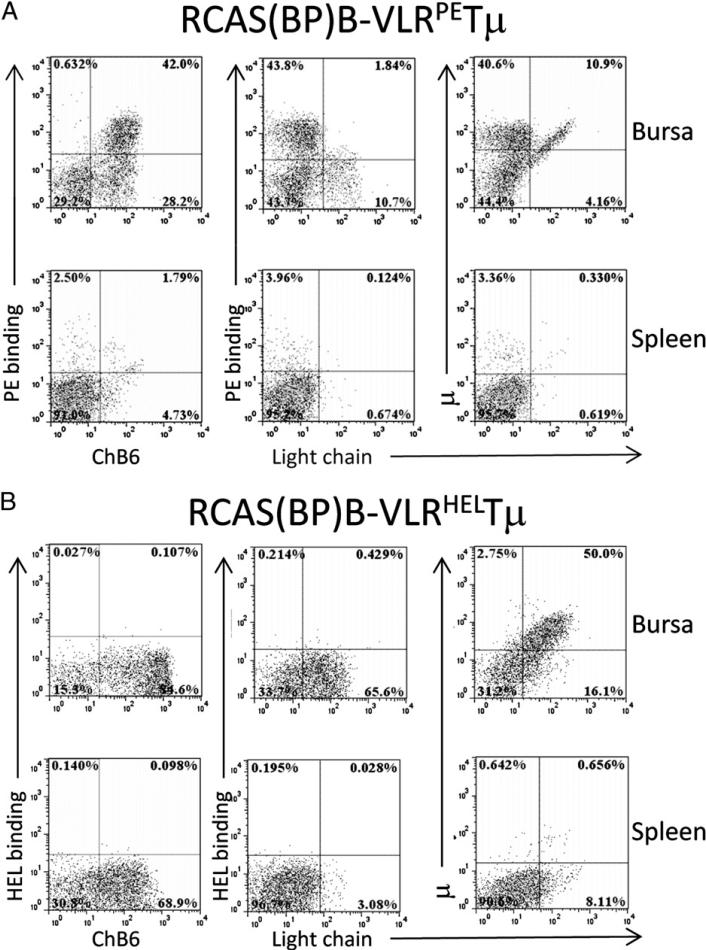
FIGURE 2.
Colonization of bursa and spleen by VLR:Tμ constructs. Bursa and splenic cells were isolated from neonatal chicks infected at day 3 of embryogenesis with RCAS(BP)B–VLRPETμ (A) or RCAS (BP)B–VLRHELTμ (B) transduced CEFs. Presence of VLRPETμ- or VLRHELTμ-expressing cells was assessed by flow cytometry on cells stained for ChB6, μ, Ig L chain, and PE or HEL. Plots are representative of 30 animals from 3 independent experiments. Dot plots are gated on forward scatter and side scatter, and 50,000 cells are represented.

