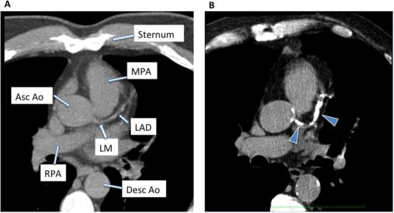
Figure 1.
Examples of noncontrast computed tomography of the chest in a subject with no coronary calcium (A) and a subject with significant coronary calcification (arrowheads) seen in the left main (LM) coronary artery and left anterior descending (LAD) artery (B). Other structures shown include the main pulmonary artery (MPA), ascending aorta (asc ao), right pulmonary artery (RPA) and descending aorta (desc ao).

