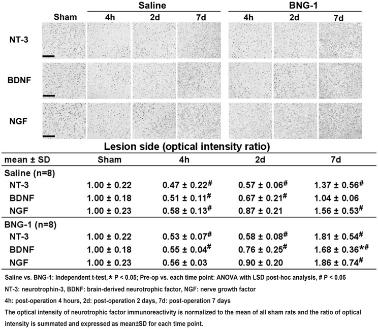
Figure 5. Comparison of the temporal expression of neurotrophic factor immunoreactivity after focal cerebral ischemia between the saline and BNG-1 treatments.
The optical intensity of BDNF immunoreactivity on the lesion cortex is higher in the BNG-1 group than the saline group at 7 d, whereas no difference in NT-3 and NGF levels occurred at any time point. The bar in the sham panels indicates 100 µm.

