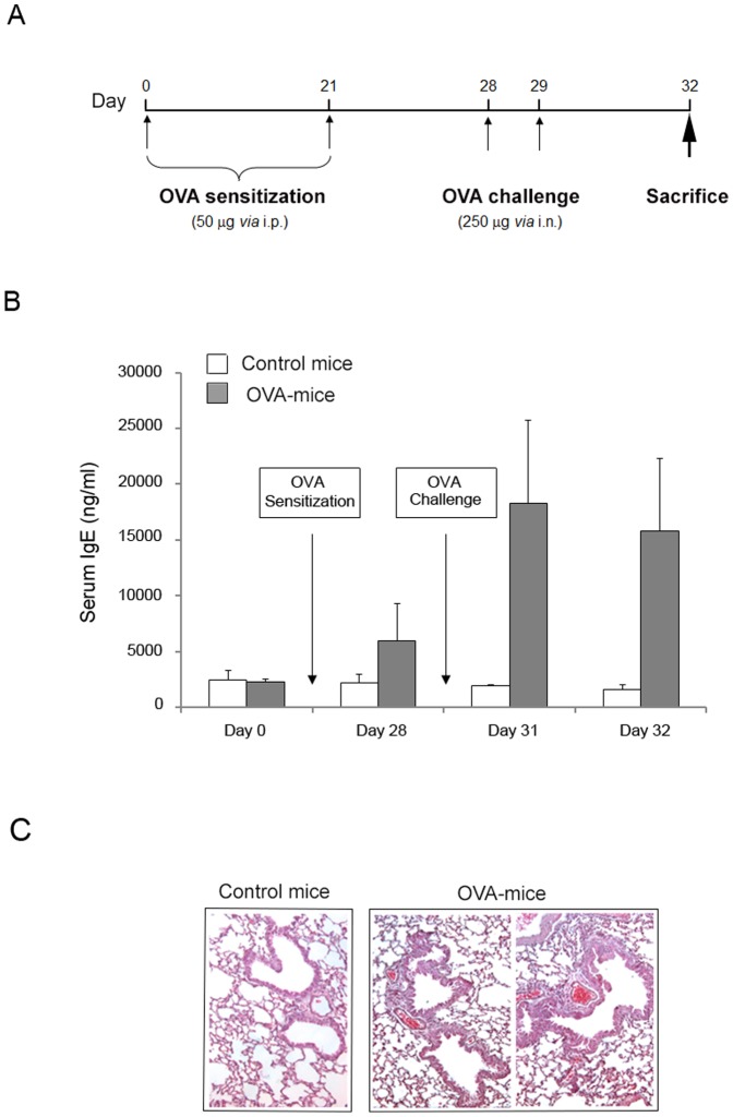
Figure 2. Establishment of airway inflammation mouse model.
Mice were exposed to an OVA “sensitization plus challenge” protocol. In A, the schedule and the timeline of OVA treatments of BALB/c mice are shown. In B, the circulating levels of total IgE were determined in serum samples of controls (n = 16) and OVA-treated mice (n = 16), harvested at the indicated experimental time points. Results are expressed as mean ± SD. In C, representative hematoxylin and eosin stained sections of lung tissue isolated (at day 32) from either healthy control or OVA-treated mice. Lungs of OVA-treated mice exhibit airway remodeling and thickness of epithelial cells (original magnification 40X).

