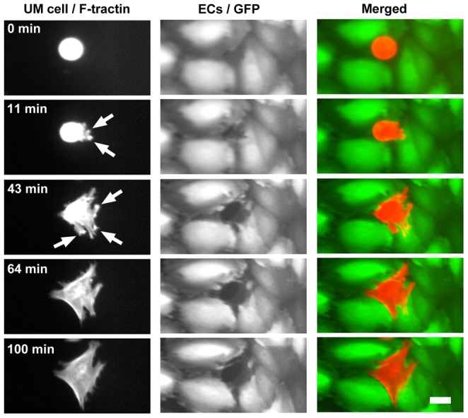
Figure 2. Invasive projections are rich in F-actin.

Frames from S2 Movie show a UM cell (OCM-1A) with F-tractin-labeled actin filaments intercalating between ECs in a monolayer. Arrows indicate F-actin-rich protrusions that transiently probe the space under the monolayer. The UM cell completes intercalation and maintains contact with adjacent ECs. Scale bar = 20 µm.
