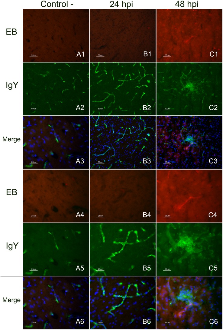
Figure 2. Immunofluorescence staining to detect IgY leakage in EB perfused and fresh frozen brain sections.
Detection of EB and IgY extravasion in the telencephalic pallium (Pall) of infected chickens at 24 and 48 hpi in comparison with a control chicken at 48 hpi (figures in the top, from A1 to C3 measure 50 µm and figures in the bottom measure 25 µm). EB extravasation (red colour) in the telencephalic pallium (Pall) was only observed in brain samples of chickens evaluated at 48 hpi (C1, C4). Images at two different magnifications showing a microvessel with a fan-like area of EB leakage (C1, C4). No EB extravasation was observed in non-infected control chickens perfused with EB at 48 hpi (A1, A4), nor EB extravasation was detected on infected chickens at 24 hpi (B1, B4). Leakage of the serum protein IgY (C2, C5) was observed in the vessels and the nearest brain cells in infected chickens perfused at 48 hpi (green colour). IgY staining in control (A2, A5) and infected chickens at 24 hpi (B2, B5) was limited to the lumen of the vessels. Merged image allowed demonstrating the presence of colocalization of IgY leakage in areas of EB extravasation in chickens evaluated at 48 hpi (C3, C6). Controls chickens evaluated at 48 hpi and infected chickens at 24 hpi did not show EB leakage and the IgY staining was limited to the lumen of the vessels.

